Le laboratoire de Neurochirurgie de la base du crâne et chirurgie cérébro-vasculaire, est destiné à faire progresser les techniques chirurgicales en Neurochirurgie et à étudier et tester de nouvelles stratégies chirurgicales.
L’anatomie du cerveau et de la base du crâne est la plus complexe de l’organisme. Une connaissance approfondie en 3 dimensions de cet environnement extrêmement complexe est un préalable indispensable à la pratique de cette chirurgie de pointe.
Cette connaissance permet au Neurochirurgien de prédire la position des structures à risques, anticiper et améliorer l’exécution de ses gestes.
L’environnement de notre laboratoire permet de recréer les conditions d’une intervention chirurgicale et donc de simuler, développer et tester de nouvelles techniques chirurgicales.
Il permet également un entrainement aux gestes minutieux et millimétrés nécessaires à la pratique de la neurochirurgie dans ces régions complexes.
La publication de nos travaux effectués dans notre laboratoire dans des revues de Neurochirurgie, destinées à la communauté neurochirurgicale, permet de diffuser ces nouvelles techniques et stratégies.
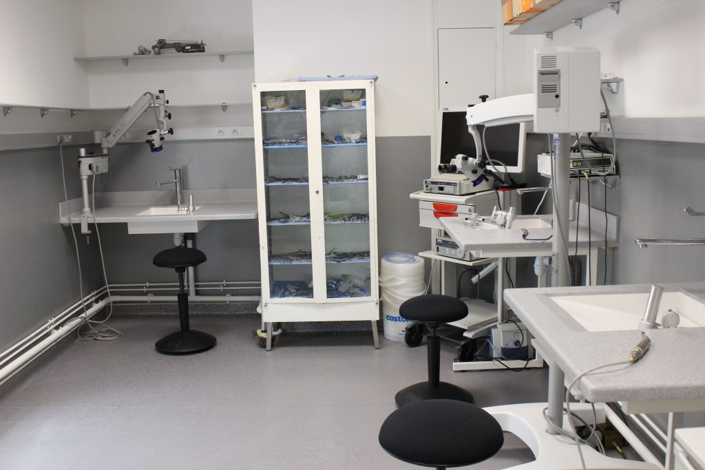
Stations de travail
Le laboratoire dispose de 4 stations de travail avec pour chaque station un équipement chirurgical comparable à celui d’une salle d’opération,
Cours de formation
Notre laboratoire permet l’organisation, plusieurs fois par an, de cours pratiques de formation de haut niveau, destinés à des neurochirurgiens souhaitants se perfectionner dans ces techniques de pointe.
Équipements du laboratoire
- Microscope opératoire- Zeiss
- Endoscope – Storz
- Colonne vidéo HD- Storz
- Station de Neuronavigation Medtronic
- Moteur haute vitesse – Medtronic
- Mayfiel head holder – Integra
- Instruments de microchirurgie
- Modèle pour anastomose vasculaire
L’équipement est mis à notre disposition par nos sponsors :
- ZEISS
- STORZ
- INTEGRA
- MEDTRONIC
Fellows et projets de recherche précédents
Dr Francesco Corrivetti, Italie : Techniques endoscopiques appliquées à la chirurgie insulaire.
Dr Kenishi Oyama, Japon : V1-V2 anterolateral triangle.
Dr Wei Chieh CHANG, Taishung, Taiwan : Transorbital endonasal approach to the temporal lobe.
Dr Shunya Hanakita, Tokyo, Japon : Deep skull base lesions : Multiportal –multimodal approach.
Dr Hun-Ho Park, Seoul, Corée : Endoscopic endonasal approach to the mesial temporal lobe.
Dr Daniel Ranconi, Belohorizonte, Brésil : Trans-orbital approach to the mesial temporal lobe.
Dr Valentino Tardivo, Turin, Italie: Endoscopic assisted approach to the craniocervical junction through the far lateral approach.
Dr François Rhéault, Canada
Dr Miguel Marigil Sanchez, Madrid, Espagne: Comparative study of the transorbital and supraorbital approaches.
Dr Davide Di Carlo, Pisa, Italie: Endoscopic assisted approach of the clivus using the anterolateral approach for the craniovertebral junction.
Dr Calvin Mak, Hong-Kong : Transorbital approach
Dr Steven Hsu, Taïpe, Taiwan: Transorbital versus supra-orbital approach
Dr Danilo Malta, Brazil: Combined petrosal approaches and the transposition of the TS-SS junction.
Dr Bremo Camara, Belo-horizonte, Brazil, sujet de recherche: Inferior eyelid approach to the mesial temporal lobe
Dr Fumihiro Matano, Tokyo, Japan: Vertebral artery transposition : classification and technique
Dr Atsuchi Okano, Tokyo, Japan: Prelacrymal transmaxillary endoscopic approach to the lateral skull base
Dr Arianna Fava, Pisa, Italy: Minicombined transpetrosal approach
Dr Tancredo Alcantara, Belo horizonte, Brésil
Dr Jerold Ness, Manille, Philippines
Publications
Endoscope-assisted anterolateral approach to a recurrent cervical spinal chordoma.
How I do it: combined petrosectomy.
Distance Control and Virtual Drilling Improves Anatomical Orientation During Anterior Petrosectomy.
The Chopsticks Technique for Endoscopic Endonasal Surgery-Improving Surgical Efficiency and Reducing the Surgical Footprint. Labidi M, Watanabe K, Hanakita S, Park HH, Bouazza S, Bernat AL, Froelich S. World Neurosurg. 2018 Sep;117:208-220.
Endoscopic Endonasal Approach to the Anteromedial Temporal Fossa and Mobilization of the Lateral Wall of the Cavernous Sinus Through the Inferior Orbital Fissure and V1-V2 Corridor: An Anatomic Study and Clinical Considerations. Hanakita S, Chang WC, Watanabe K, Ronconi D, Labidi M, Park HH, Oyama K, Bernat AL, Froelich S. World Neurosurg. 2018 Aug;116:e169-e178.
Combined Nasoseptal and Inferior Turbinate Flap for Reconstruction of Large Skull Base Defect After Expanded Endonasal Approach: Operative Technique. Boetto J, Labidi M, Watanabe K, Hanakita S, Bouazza S, Passeri T, Bernat AL, Froelich S. Oper Neurosurg (Hagerstown). 2019 Jan 1;16(1):45-52.
Endoscopic Approach of the Insula Through the Anterior Middle Temporal Gyrus: A Feasibility Study in the Laboratory.Corrivetti F, Froelich S, Mandonnet E. Oper Neurosurg (Hagerstown). 2017 Jul 25.
Surgical Anatomy for the Endoscopic Endonasal Approach to the Ventrolateral Skull Base. Oyama K, Tahara S, Hirohata T, Ishii Y, Prevedello DM, Carrau RL, Froelich S, Teramoto A, Morita A, Matsuno A. Neurol Med Chir (Tokyo). 2017 Oct 15;57(10):534-541.
Paratrigeminal, Paraclival, Precavernous, or All of the Above? A Circumferential Anatomical Study of the C3-C4 Transitional Segment of the Internal Carotid Artery. Marcati E, Andaluz N, Froelich SC, Zimmer LA, Leach JL, Fernandez-Miranda JC, Kurbanov A, Keller JT. Oper Neurosurg (Hagerstown). 2018 Apr 1;14(4):432-440.
Drilling of the marginal tubercle to enhance exposure via mini pterional approach: An anatomical study and clinical series of 25 sphenoid wing meningiomas. Aldahak N, El Tantowy M, Dupre D, Yu A, Keller JT, Froelich S, Aziz KM. Surg Neurol Int. 2016 Dec 12;7(Suppl 40):S989-S994.
Neuronavigated Fiber Dissection with Pial Preservation: Laboratory Model to Simulate Opercular Approaches to Insular Tumors. Mandonnet E, Martino J, Sarubbo S, Corrivetti F, Bouazza S, Bresson D, Duffau H, Froelich S. World Neurosurg. 2017 Feb;98:239-242.
The Medial Extra-Sellar Corridor to the Cavernous Sinus: Anatomic Description and Clinical Correlation. Theodosopoulos PV, Cebula H, Kurbanov A, Cabero AB, Osorio JA, Zimmer LA, Froelich SC, Keller JT. World Neurosurg. 2016 Dec;96:417-422
Segments of the internal carotid artery during endoscopic transnasal and open cranial approaches: can a uniform nomenclature apply to both? DePowell JJ, Froelich SC, Zimmer LA, Leach JL, Karkas A, Theodosopoulos PV, Keller JT. World Neurosurg. 2014 Dec;82(6 Suppl):S66-71.
Endoscopic, endonasal variability in the anatomy of the internal carotid artery. Cebula H, Kurbanov A, Zimmer LA, Poczos P, Leach JL, De Battista JC, Froelich S, Theodosopoulos PV, Keller JT. World Neurosurg. 2014 Dec;82(6):e759-64.