
Symptoms
Chordomas grow slowly, and symptoms are generally late. They vary depending on the location:
Skull base
Headaches, double vision, loss of vision or hearing, difficulty swallowing, hoarseness, gait disturbances, motor weakness.Spine
Pain, tingling, or a feeling of weakness in a limb.Sacrum
Often discovered late: pain, urinary disorders, or even urinary and fecal incontinence.Diagnosis
CT and, above all, MRI can identify the tumor. The very characteristic appearance of chordomas on MRI often makes it possible to strongly suspect the diagnosis. Specific MRI sequences are required—because this is a rare tumor, radiological diagnosis requires experienced radiologists.
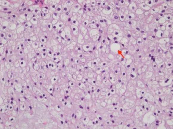
Types of chordomas
The 2020 WHO classification defines three categories:
-
Conventional chordoma
Includes typical and chondroid chordomas. Represents the majority of cases.
-
Dedifferentiated chordoma
Very rare, more aggressive. Most often occurs during the course of a known chordoma, particularly in the event of recurrence after irradiation.
-
Poorly differentiated chordoma
A new entity defined by loss of the INI1 protein (deletion of the SMARCB1 gene). More aggressive than conventional chordoma, but may respond to targeted medical treatments.
Treatment
Multidisciplinary approach
A multidisciplinary approach is essential. Our department organizes a monthly national MDT meeting “Chordomas and Chondrosarcomas”, enabling all French teams to present their cases and discuss therapeutic options. We collaborate in particular with:
- The Neuroradiology Department at Lariboisière Hospital
- The Orsay proton therapy department (Institut Curie — Dr Hamid Mammar, Dr Valentin Calugaru)
- Necker Hospital — pediatric neurosurgery department (Prof. Kevin Beccaria)
- Biopsy Rarely necessary at the skull base and cervical spine. At the lumbar level or in the sacrum, it may be performed to confirm the diagnosis—always by trained teams, under strict conditions to avoid tumor seeding along the biopsy tract.
- Surgery Must be performed in experienced centers. Incomplete treatment in an insufficiently specialized center can compromise any subsequent surgical reoperation. The goal is complete resection while preserving neural structures. At the sacrum, en bloc resection (without crossing the tumor capsule) is the optimal technique to maximize the chances of cure.
- Proton therapy / Radiotherapy Chordomas have a strong tendency for local recurrence—surgery must therefore be complemented by high-dose radiotherapy. The reference technique is proton therapy (Orsay center). Radiosurgery may be offered for small-volume lesions or small localized recurrences.
- Chemotherapy In the event of recurrence, reoperation is most often proposed first. If a further recurrence occurs or if surgery is not possible, chemotherapy may be considered. Clinical trials are underway to evaluate new treatments.
Course and prognosis
The course is often favorable but requires aggressive treatment combining surgery and proton therapy. Each patient’s prognosis is unique and depends on age, the type of chordoma, tumor volume at diagnosis, location, and whether resection is complete.
Approximately 30% of patients develop metastases during the course of the disease, most often late (lungs, liver, subcutaneous tissue, lymph nodes). They are rarely present at the initial diagnosis and are often well tolerated.
Useful links

Chordomas develop from residues of the notochord. The notochord is an embryonic structure that forms the axis of development of the fetus and participates in the development of the spinal column.
The notochord disappears when the fetus is around 8 weeks old, but some notochord cells do not disappear and persist in the bones of the spine and skull base. The reason why some of these cells develop into chordomas is not yet known, but research is progressing.
The vast majority of chordomas occur randomly and are not hereditary. As with all tumors, there are certain genetic changes in chordoma cells.
For example, over 95% of individuals with a chordoma have an abnormality in the DNA sequence of a gene called brachyuria. This anomaly increases the risk of developing a chordoma, but does not in itself cause a chordoma.
There are around 80 new cases in France every year. Indeed, at any given time, fewer than one person in 100,000 is living with a chordoma (prevalence).
Chordoma is most often diagnosed in people aged 50–60, but it can occur at any age.
In addition, there is a slight male predominance, with a ratio of about 2 men to 1 woman.
Familial chordomas are very rare, and only a few familial cases have been identified.
Around 40% of chordomas form in the skull base and craniocervical hinge, 30% in the bones of the mobile spine and around 40% in the sacrum.
Chordomas of the skull base are generally located in a bone called the clivus. These are deep-seated lesions, generally located in the center of the skull, close to critical structures, which are difficult to access and complex to treat. Skull base chordomas are sometimes called brain tumors because they can grow inwards from the skull towards the brain, but they don't actually develop from brain cells.
They can also occur in the cervical spine and lumbar spine (lower back). These zones make up the "mobile" column. Locations in the thoracic spine are rarer.
In 40% of cases, they also occur in the sacrum. They are located more or less high on the sacrum and are more or less complex to remove, depending on the relationship with the nerves to the sphincters and the rectum in front.