 English
 Français
Cerebral vascular malformations are congenital or acquired lesions consisting of vessels and localized in the brain or on the surface. They may manifest as intracerebral or pericerebral (subarachnoid) haemorrhage. Some may be serious in case of bleeding, others have a very low risk of bleeding and are therefore benign. Several therapeutic modalities are possible (surgery, endovascular treatment inside the vessels, radiotherapy) according to the size, the configuration of the malformation and the general state of the patient. These modalities are discussed in multidisciplinary consultation in order to offer the optimal therapeutic solution to each patient.
Type vascular malformation of the brain
We distinguish :
- aneurysms
- arteriovenous malformations (AVM)
- dural arteriovenous fistulas (AVf)
- cavernomas
- venous angiomas
- telangiectasias
How do they manifest?
All these vascular malformations have in common the fact that they can manifest themselves brutally by a haemorrhage:
- bleeding into the subarachnoid space around the brain which is called a subarachnoid haemorrhage. It is most often due to an aneurysm rupture
- bleeding into the cerebral parenchyma which is called a cerebral or cerebellar hematoma. It is most often due to the rupture of an AVM or a cavernoma. Intra-cerebral hematoma can also have other causes including trauma, rupture of high blood pressure, tumor… It can be favored by anticoagulant or an antiplatelet drug.
- cerebro-meningeal hemorrhage when the bleeding occur in the subarachnoid space and in the brain.
All these vascular malformations also have another common point: they can be accidentally discovered during a CT scan or an MRI performed to look for something else. Finally, they can also present with seizure (epilepsy).
Therapeutic modalities used in vascular neurosurgery
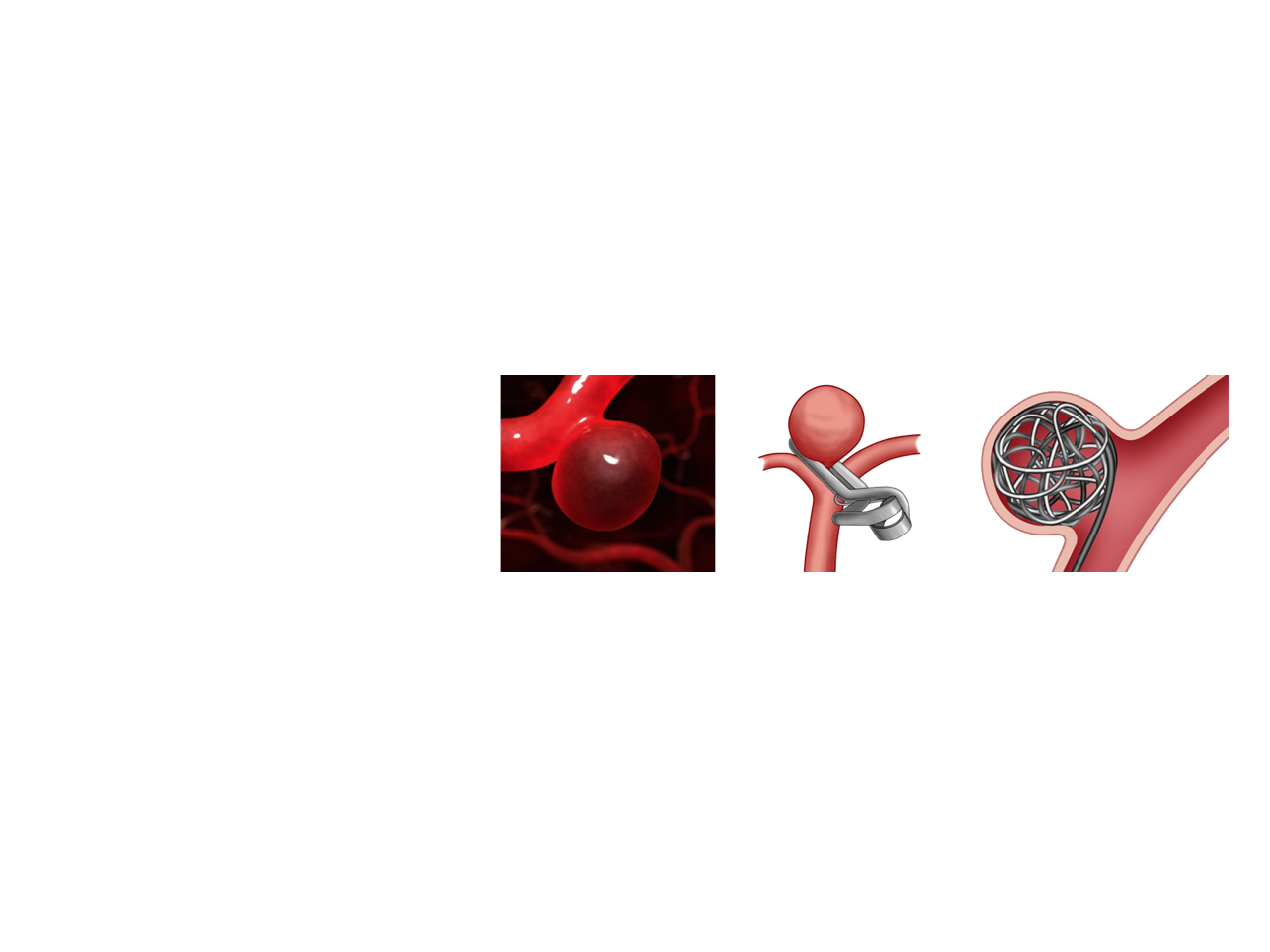
- Surgery
Surgical exclusion of aneurysm or arteriovenous malformation requires opening of the skull. In the case of aneurysms, after isolating the aneurysm sac and separating the important arterial branches surrounding it, a clip (sort of micro-“clothespin”) is applied on the base of the aneurysm to exclude the aneurysm of the blood circulation. This prevents the rupture of the bag or its re-bleeding in case of aneurysm already broken.
- Embolization
Embolization of aneurysm, arteriovenous malformation, or dural fistula is a procedure for occluding the vascular malformation without opening the skull. The malformation is accessed through the interior of the vessels. It consists of the insertion of microcatheters by puncture of the femoral artery (artery of the leg). The catheter is brought to the malformation following the vessels. The progression of the catheter to the malformation is followed on screens by the injection of visible contrast material on X-rays performed in real time. Once the catheter is in the malformation, the malformation is occluded by the injection or placement in the malformation of different materials. Several devices can be set up in the malformation by this method, such as coils (metal microspires), stents, microparticles or glue. The goal is to prevent blood flow from circulating through the vascular malformation by filling it with the chosen material and / or by blocking access to the blood stream (stent).
- Radiosurgery
Radiosurgery is a technique of radiation therapy. It is most often used for brain tumors, but can also be applied to other areas of neurosurgery, including the treatment of arteriovenous malformations. The trajectory of the radiation is precisely calculated to reach only the target without irradiating the healthy brain around the malformation. This technique requires the installation of a frame attached to the head under local anesthesia or the application of a face mask depending on the technology used. This is a painless procedure, performed as an outpatient and requiring one to a few sessions.
- Bypass
Bypass in neurosurgery is the equivalent of coronary bypass surgery in cardiac surgery. It involves connecting two arteries by micro-sutures to revascularize a territory of the brain. It is a technique more rarely needed for the treatment of Moya-Moya disease, the treatment of complex aneurysms or the revascularization of insufficiently perfused brain areas. Bypassing may also be necessary when a large tumor-infiltrated brain vessel must be sacrificed to allow removal of the tumor.
The neurovascular medical-surgical team
Each patient presenting a surgical neurovascular pathology is discussed during multidisciplinary meetings in the presence of the neurosurgery team (Pr. Froelich department), the interventional neuroradiology team (Pr. Houdart department), the team of neurology (Dr. D. Hervé). A therapeutic decision is made according to the specificities of each patient and his situation.
Research and training
The departments of Neurosurgery, Interventional Neuroradiology and Neurology are developing research and training activities in various fields of neuromuscular diseases in order to better understand the origin and developmental mechanisms of aneurysms and AVMs and to improve patient care.
The CERVCO
The Neurosurgery Department is part of the CERVCO, a specialty group specializing in the management of rare vascular diseases of the retina, brain and spinal cord.
The missions of CERVCO are multiple:
• Improve the management of rare diseases in relation to all clinicians at the national level and in coordination with the family associations concerned,
• Establish protocols for the management of these conditions and adapt them regularly according to changes in research,
• Disseminate in a general way all the useful information to the patients, to their families, to all the doctors or carers taking care of these pathologies at the national level.
• To improve the knowledge of the mechanisms of these affections thanks to the research developed within CERVCO, because this is essential for the development of new therapies.

![]()